Sildomar Queiroz E Silva 1, Sabrina Ramos Biano 2, Daniella Paula Dias Coelho 3, Felipe Pereira de Loredo 4
Resumo
Patient asymptomatic in a mammographic screening exam where no lesions suspected of malignancy were seen. A tomosynthesis was performed where a spiculated nodule in the right breast.Dados do caso
Feminino, 48 anos.
Palavras chaves
Mammography, Breast Neoplasms.
Histórico Clínico
Female patient, 48 years old, with no family history of breast cancer, asymptomatic in a mammographic screening exam where no lesions suspected of malignancy were seen. A tomosynthesis was performed where a spiculated nodule in the right breast with BI-RADS V classification was identified. The histopathological examination showed invasive ductal carcinoma.
Achados Radiológicos
A mammographic screening exam where no lesions suspected of malignancy were seen. A tomosynthesis was performed where a spiculated nodule in the right breast with BI-RADS V classification was identified.
Discussão
Breast cancer is the second most common neoplasm of women in Brazil, second only to non-melanoma skin cancer. Data from the National Cancer Institute (Inca) estimated about 59,700 new cases of breast cancer in women in the year 2018 and each year there is an expectation of a 29% increase in new cases in the country (1,2). The main risk factors are related to gender, age, heredity, endocrine and behavioral factors, such as alcoholism and smoking. Among those, the most relevant are inserted in the context of genetic / hormonal changes and about 75% of cancer cases are related to them and are difficult to detect (3). In this context, preventive actions are essential for the diagnosis and early treatment of breast neoplasm, and mammography is the imaging method considered a gold standard for screening with an effective capacity to reduce the risk of death by the disease by up to 25% . Mammography has been evolving and complementing other imaging methods over the years in order to allow greater diagnostic efficacy. The factors related to this performance are related to the technique and technology employed as well as the composition of the studied breast tissue, losing expressive sensitivity in breasts of higher density. In these cases, other imaging methods allow better study of breast tissue such as ultrasonography and more currently tomosynthesis (4,5). The tomosynthesis allows the acquisition of three-dimensional images being able to overcome some limitations of conventional mammography, such as tissue overlapping that may mask some lesions or even mimic non-existent lesions (6,7,8) . Some studies, such as that by Nicholson, et al., suggest tomosynthesis as a surrogate method to conventional mammography and that better target complementary examinations, such as ultrasonography and resonance. Other studies also show that the sum of tomosynthesis and mammographic screening has reduced the reconvocation of patients to complement new images in cases of doubtful lesions at diagnosis, that is, they increase the accuracy of detection of these images (9,10). In this report we present a case of breast lesion that is best visualized in the tomosynthesis.
Lista de Diferenciais
Diagnóstico
Aprendizado
In this clinical case, a precise definition and identification of lesion borders and visualization of the spiculated margins was possible with tomosynthesis, not seen previously on mammography screening. The precise definition by the reduction of the tissue overlap factor that can occur in the conventional mammography can be a useful tool for the evaluation.
Referências
1. Instituto Nacional de Câncer José Alencar Gomes da Silva. Incidência de Câncer no Brasil. Disponível em: http://inca.gov.br/estimativa/2018/. Acesso em 18 de Abril de 2018.
2. Ohl ICB, Ohl RIB, Chavaglia SRR, Goldman RE. Public actions for control of breast cancer in Brazil: integrative review.Rev Bras Enferm [Internet]. 2016;69(4):746-55. DOI: http://dx.doi.org/10.1590/0034-7167.2016690424i
3. Silva P A, Riul S S. Câncer de mama: Fatores de risco e detecção precoce. Rev Bras Enferm, Brasilia, 2011; 64 (6): 1016-21.
4. Rosa LM,Silva L, Radunz V, Anzuaga MA. Rastreamento mamografico: Detecção de lesões neoplásicas malignas em mulheres de Santa Catarina e do Brasil. Texto contexto Enferm., 2016; 25(3); e 5280015.
5. Villar VCFL, De Seta MH, Andrade CLT, Delamarque EV, Azevedo ACP. A evolução da qualidade da imagem em mamografia no Estado do Rio de Janeiro.Radiol Bras. 2015 Mar/Abr;48(2):86–92.
6. Vilaverde F, Rocha A, Sousa M R, Mesquita R, Reis A. Tomossíntese mamária: O que o radiologista deve saber. Acta Radiológica Portuguesa. 2016, n° 109, volume XXVIII: 35-41.
7. Nascimento FB, Pitta MGR, Rêgo MJBM. Análise dos principais métodos de diagnóstico de câncer de mama como propulsores no processo inovativo. Arquivos de medicina 2015;29(6):153-159.
8. Seabra Z T, Lourenço J. Imaginologia no carcinoma da mama. Revista Portuguesa de Cirurgia (2013) (27):59-70.
9. Peppard HR, Nicholson BE, Rochman CM, et al. Digital Breast Tomosynthesis in the Diagnostic Setting: Indications and Clinical Applications. Radiographics. 2015 Jul-Aug;35(4):975-90.
10. Chesebro AL, Chikarmane SA, Ritner JA, Birdwell RL, Giess CS. Troubleshooting to Overcome Technical Challenges in Image-guided Breast Biopsy. Radiographics. 2017 May-Jun; 37(3):705-718.
Imagens
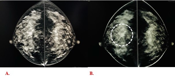
Figure 1:
A- Digital mammography synthesized in the right and left cranial caudal views.
B- Tomosynthesis in the right and left cranial caudal views, showing spiculated lesion.
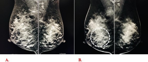
Figure 2:
A- Digital mammography synthesized in the right and left medial lateral views.
B- Tomosynthesis in the right and left lateral median views, showing spiculated lesion.
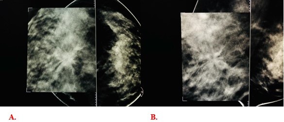
Figure 3: Magnified image of spiculated lesion visualized on tomosynthesis in the cranial caudal (A) and lateral medial (B) views.
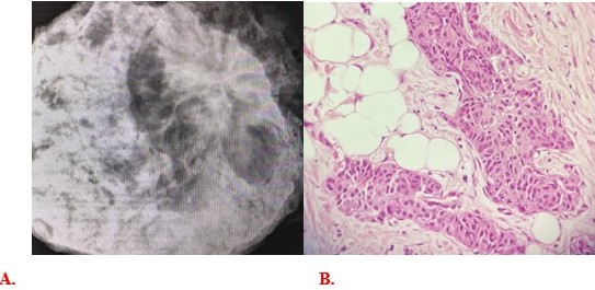
Figure 4:
A- Peça cirúrgica radiografada demostrando lesão espiculada.
B- Histopathology show invasive ductal breast carcinoma.
Artigo recebido em domingo, 1 de março de 2020
Artigo aprovado em quarta-feira, 15 de abril de 2020
 Todos os artigos científicos publicados em brad.org.br são licenciados sob uma licença Creative Commons.
 Português PDF
 Imprimir
 Envie este artigo por e-mail
 Como citar esse artigo
 Envie um comentário
 Mendeley
 Pocket