Arquimedes Artur Zorzetto 1, Flávia Sprenger 2, Karin Cristiane Ferreira 3, Maria Cristina Sartor 4
Abstracto
We report a case of a 42-year-old male patient with a history of Crohn's disease that presented to a barium small bowel study with a rare type of Phytobezoar. This case aims to review epidemiological and imaging aspects of seed bezoars.Dados do caso
Masculino 42 anos
Palavras chaves
Doença de Crohn, Sulfato de Bário, Tomografia
Histórico Clínico
A 42-year-old male patient with a known history of Crohn’s disease presented to the Radiology department for a small bowel barium study. The patient was in clinical remission, under no treatment for the past four years. His latest colonoscopy showed an ileal stenosis.
Achados Radiológicos
Plain abdominal radiograph showed three oval radiopaque images with a less dense center on his lower left abdomen (figure 1). These same images were also seen on a lumbar spine radiography performed a year earlier. When barium reached the distal ileum, the previously described images presented as filling defects, proving to be inside the small intestine’s lumen. Adjacent to filling defects, the patient had two significant stenosis, right before and after them (figure 1). The images moved freely inside the small space between the focal stenosis during compression and by changing the patient’s position. One year later the patient underwent a computed tomography enterography that showed signs of chronic ileitis, and the same bezoars remained on the same small bowel segment (figure 2).
Discussão
At the end of the study the patient was questioned about possible seed ingestion, but he did not recall any events. Since a phytobezoar was the most plausible hypothesis, the attending radiologists radiographed common fruits consumed in South Brazil, such as jaboticaba (Myrciaria cauliflora), plum (Prunus domestica), araticum (Annona coriacea) and pitanga (Eugenia flora) (figure 3). None of them, however, had a radiological aspect similar to the patients’ images. A professor of seed technology was consulted, and he suggested that the images had the radiological aspect of a palm tree seed, possibly a pupunha seed (Bactris gasipaes). A pupunha seed was then radiographed and was identical to the patient’s assumed bezoar (figure 4). This seed has a rigid cover and a soft center, unlike most seeds. Few cases of seed bezoars have been reported in the literature and even less on patients with Crohn’s disease. Watermelon and sunflower seed bezoars dominate the case reports on the literature on hygid patients, but there is no previous report of a pupunha seed phytobezoar [1, 2, 3]. The red pupunha fruit is one of the most consumed fruits in Northern Brazil, eaten raw or cooked. They are rich in antioxidants and proved to have anti-lipidemic and inti-inflammatory properties, and also have a large impact on local industry and economy [4, 5]. Imaging played an important role on this case, especially since the patient was asymptomatic and no invasive conduct was taken to actually deduce the bezoar’s etiology. This was the main limitation of the present study. However, our goal was to emphasize the occurrence, despite of its rarity, of seed bezoars on patients with Crohn’s disease and to document the radiographic aspect of common fruits in Brazil.
Lista de Diferenciais
Diagnóstico
Aprendizado
In this case we learned to be alert to silent signs of inflammatory bowel disease. This patient was asymptomatic but presented with focal stenosis, causing the bezoars to be lodged within an intestinal loop, and other signs of disease activity despite poor clinical presentation.
Referências
Imagens
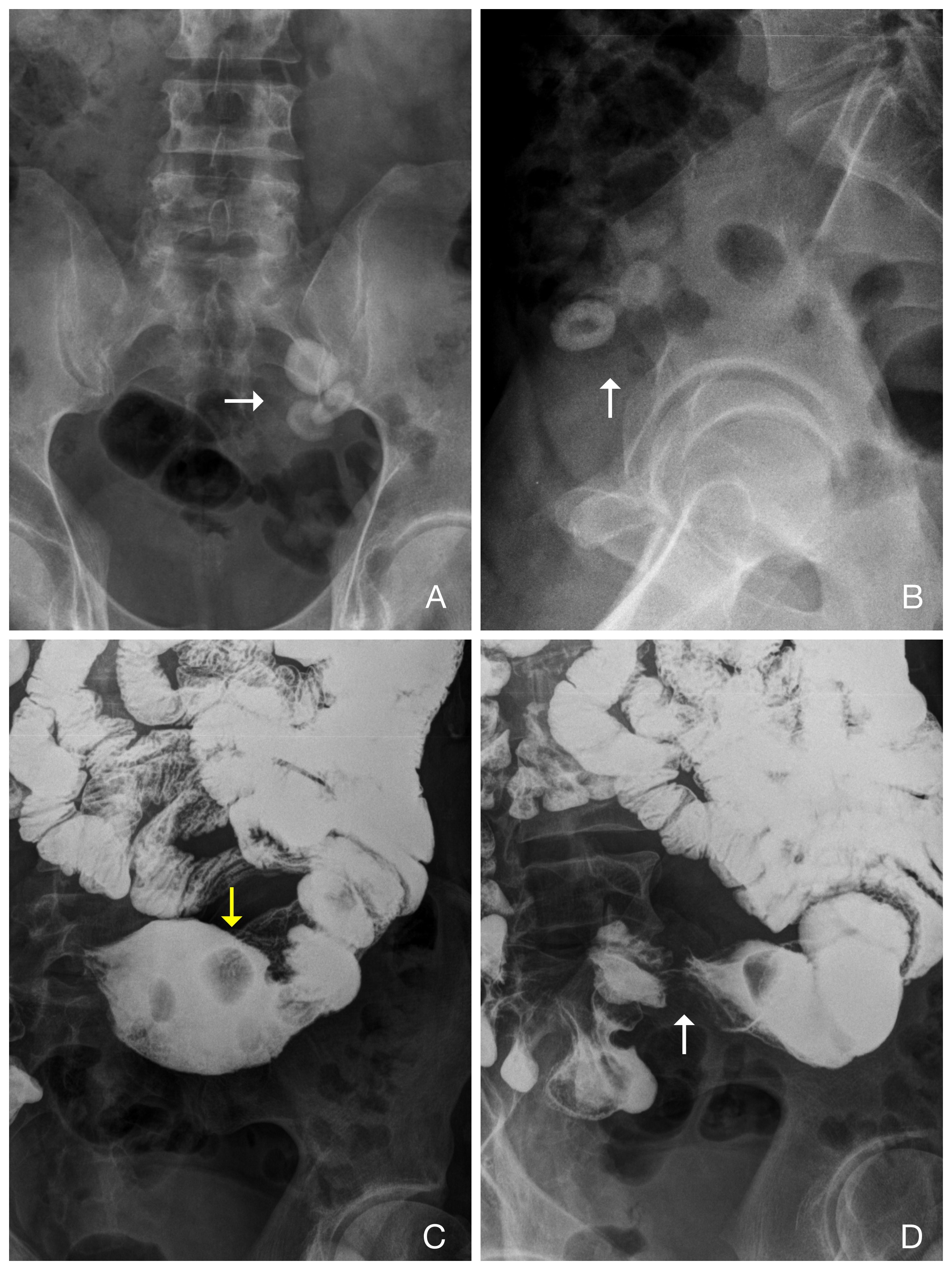
Fig. 1. A: plain abdomen radiograph (anteroposterior projection). B: Plain abdomen radiograph (profile projection). A and B: Plain abdomen radiographs reveal three small oval radiopaque images, with a less dense center, projected in the lower left abdomen (arrows). C and D: Abdomen radiograph 40 minutes after the barium administration. The previous radiopaque images proved to be inside the small intestine, appearing at this moment as filling defects (yellow arrow). During the dynamic study, with patient movement and focal compression the filling defects proved to be confined between two spots of focal stenosis (white arrows)
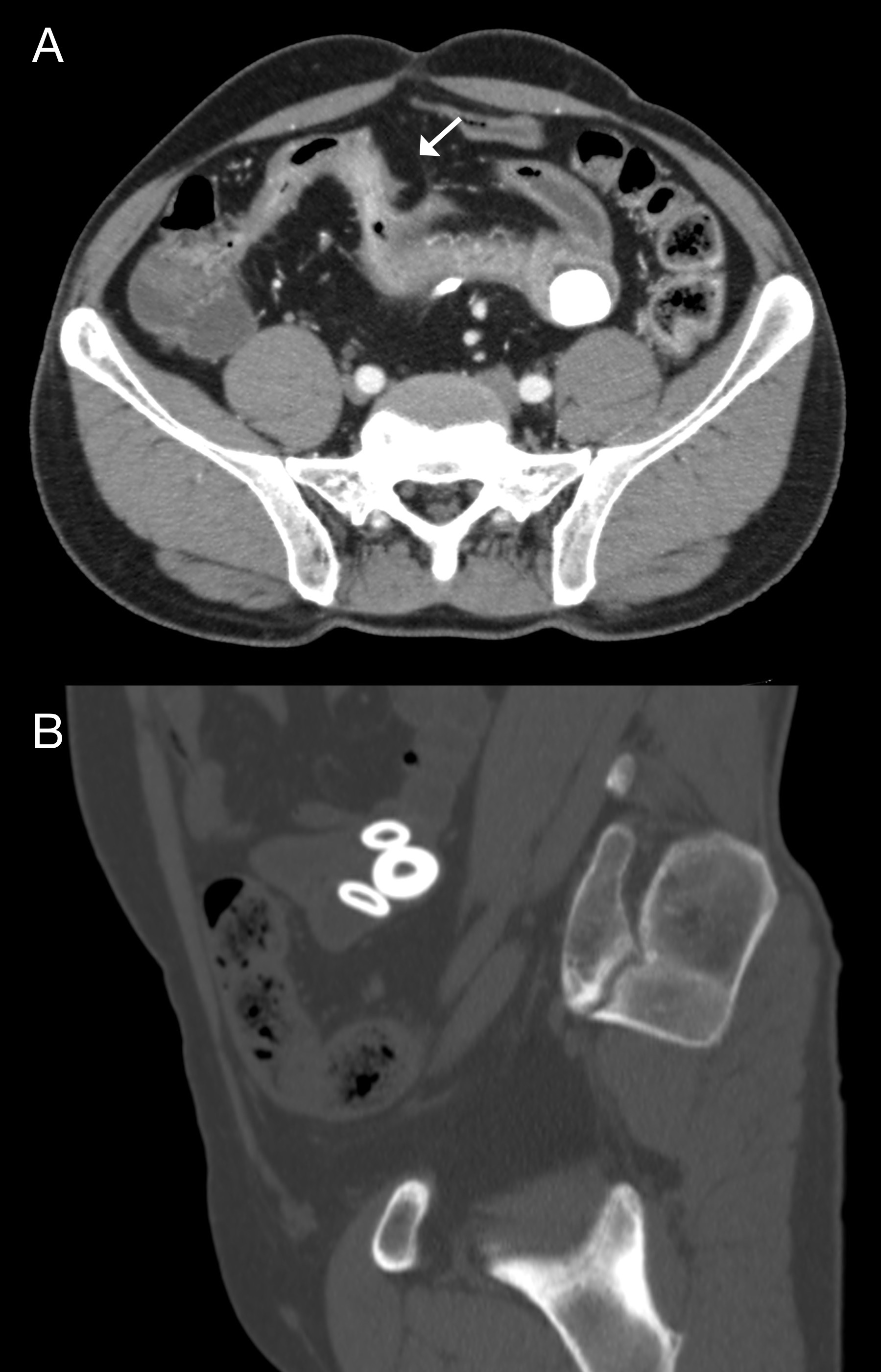
Fig 2. A: Axial image from the patient’s computerized tomography enterography showing a terminal ileal wall thickening, with increased mucosal enhancing and reduced caliber (arrow). These are all signs of active chronic ileitis. Notice the bezoar inside an intestinal loop on the left lower quadrant of the abdomen. B: Sagital imaging using bone window revealing the three bezoars remain inside the same intestinal loop one year after the barium study.
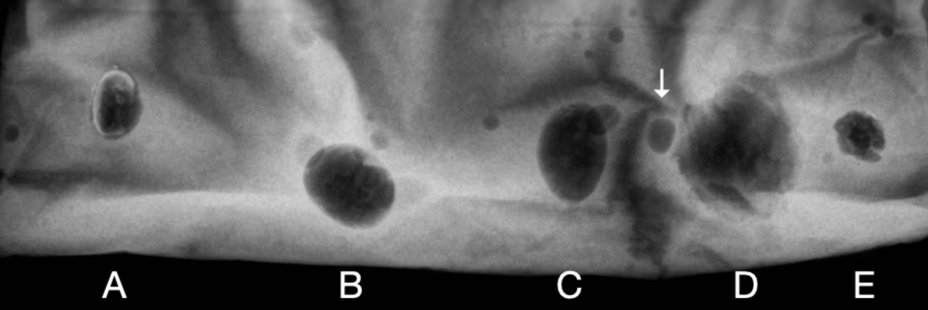
Fig. 3. Fruit seeds radiographed inside a bag filled with barium, simulating a barium small bowel study. A and B: Jabuticaba seed (Myrciaria cauliflora). C: Araticum seed (Annona coriacea). D: Plum seed (Prunus domestica). E: Pitanga seed (Eugenia flora). Notice some air bubble artifacts (arrow).
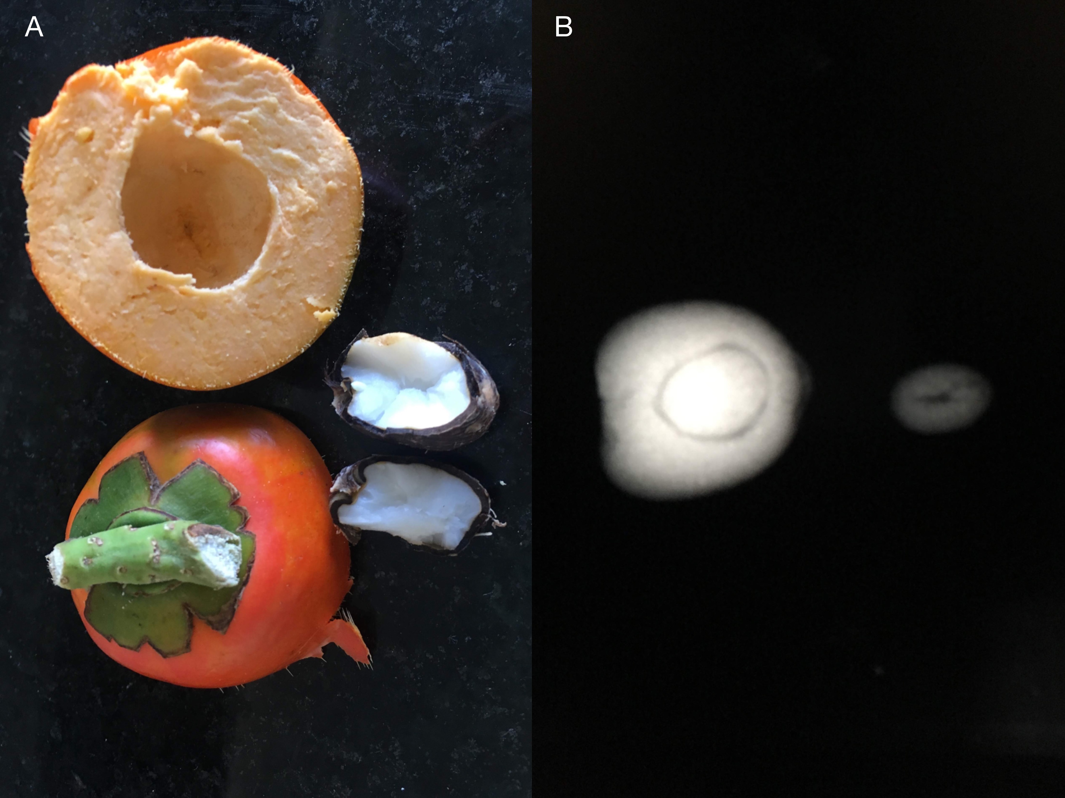
Fig. 4. A: Pupunha fruit and seed open in half, revealing a soft center and justifying its radiographic aspect. B: pupunha seed radiograph, showing a radiopaque cover and a less dense center.
Vídeos
Artículo recibido en viernes, 24 de abril de 2020
Artículo aceptado el lunes, 27 de abril de 2020
 Todos los artículos científicos publicados en brad.org.br están bajo una licencia Creative Commons.
 Portugués PDF
 Impresión
 Enviar este artículo por correo electrónico
 Cómo citar este artículo
 Enviar un comentario
 Mendeley
 Pocket